Epidermis -- Time Dimension of the Skin

WHAT IS IT?
-----------

 Think of your friend. You meet him frequently  and his appearance seems
 always the same. Yet his unchanging appearance hides a world in flux. His
 body is made of cells. Some are born, others are dying, yet the organism
 maintains the same appearance. The following model is a glimpse into this
 fascinating world of flux. It serves two additional purposes. It
 illustrates a new way to teach Histopathology (the science of cells and
 tissues in the body). Finally it highlights an ancient philosophy.

 Let's examine this artificial skin, and see how it behaves.

HOW TO USE IT
--------------

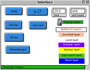
 'setup' button:             Clears the display window and creates a  blue
 			     stem cell.
 'go'  button:               Starts the simulation.
 'Create a new unit' button: Adds a new stem cell to the skin.
 'Chemotherapy' button:      Kills supra-basal cells.
 'Suntan' button:            Exposes the skin to the sun.
 'Bruise' button:            Bruises the skin.

 Two monitor tools display time and the number of skin units (unit-count)
 'Display-skin-patch' switch:  When set to 1, displays a skin patch.
 A Plot window displays the total cell count in the skin.

 The model simulates only the upper covering of the skin called epidermis.
 Its layers are displayed in the interface window. Basal layer is the
 deepest. The upper cornified covers the skin. It becomes apparent to you
 when observing the skin.

THINGS TO NOTICE
----------------

 Set 'Display-skin-patch' switch to 0.
 Click on setup. A blue skin stem cell appears.
 Click on 'Go' and watch the skin unit elongating.

 Stem cells and transitional cells.

 Only the stem cell is dividing. Following its division one of its progeny
 replaces the parent cell and remains a stem cell. The other is pushed
 upward and is called transitional cell. Stem cell division generates a cell
 stream upward.  Cells age as the go and when their time has come they die
 and are sloughed off the unit top.

 Start again. Click off the go button, then  click on setup and go. Watch
 the unit growing. As long as cell production rate exceeds the death rate,
 the unit elongates. When cell production equals  the rate of cell death,
 the unit maintains a constant length.

THINGS TO TRY
-------------

 Create a new unit (click on the so named button)

 A new stem cell is formed and it generates a new unit. Continue adding
 about 15 units. What you see is the cross section of the upper skin part
 called epidermis. Each layer has a name (given in the interface window).
 You just simulated the growth of the skin of a child. In the growing child
 the skin expands by creating additional units. Stem cells divide in two
 modes: In each unit the stem cell divides asymmetrically, since generating
 two different cells, a stem and a transitional. When a new unit is added
 the stem cell divides symmetrically, into two stem cells.  In the adult,
 stem cells do not divide symmetrically.

 Differentiation

 Cells in each layer perform specific tasks. Notice that each layer is a
 station in the upward movement of a cell. When born it is green (called
 supra basal) . Then it turns into a yellow (spinous) cell. Later it becomes
 a granular (violet) cell. At old age it turns into a cornified cell and
 then sloughs off the skin. The scales that may seed your hair, are dead
 cornified cells.
 The change in cell appaerance and its tasks is called differentiation.  Skin
 cells differentiate as they go. Notice that differentiation and aging are
 equivalent. Cell aging may be viewed as differentiation.

 Click on 'Bruise'

 When you bruise your skin, the upper layers are torn off. This condition is
 called erosion. Examine the plot window after bruising. As cells move
 upward the erosion heals. This is known also as skin regeneration.

 Click on 'Chemotherapy'

 Chemotherapy kills mainly supra basal cells. A slit of dead cells gradually
 moves upward. As long as it exists, the skin is fragile and may slough off,
 which generally does not happen. You may have heard that following
 chemotherapy the patient loses its hair. The mechanism is similar. The
 suprabasal cells of the hair follicle are killed and the hair falls out.

 Suntan

 Notice the black lines extending from the lower skin layer. These cells are
 called melanocytes. When exposed to intensive sun shine, they color all
 neighboring skin cells. Click on 'Suntan'. Repeat clicking and notice the change.
 After leaving the beach to your home, colored cells continue streaming
 upward. When they die you lose your suntan.

 Examine the skin patch.

 The model displays a microscopic cross section of the epidermis. When
 watching your skin you actually see only the upper layer. In order to watch
 the skin from above, set the 'display-skin-patch' switch to 1. Repeat the
 above experiments and watch its changing color. The real skin has a
 different color palette, yet undergoes similar changes.
 It is instructive to click on 'Suntan' and watch the patch.

 Cells stream

 What drives skin cells upward, remains a puzzle. They are neither pusheded,
 nor pulled, they simply stream.

 Now return to your friend. Although his appearance seems unchanged, you
 know that his epidermis continually changes. Not only the skin. Almost all
 cells in the body stream. They are born during a stem cell division and
 gradually stream toward their graveyard.

 Panta Rhe

 About 500 years BC a philosopher named Heraclitus said: "Everything streams
 (Panta Rhe in Greek). You never step into the same river twice". Now we may
 add, "You never meet the same person twice".


 The model

 Cell attributes (turtles-own):
 Age :
 age1:     The maximal age a cell attains
 stem :    In stem cells, stem = 1.  In transitory cells, stem = 0.
 progeny:  Each symmetric stem cell division creates a new progeny
              (setprogeny progeny + 1)
 color:    Stem cells are blue.
           In transitory cells color represents age. (setc yellow + 0.4 * age)
 melanocyte: Black cells situated among the lower layers.
 melanin:  Stores brown color. (setmelanin 35 setc melanin)

 patches-own [skin?] Displays the skin as viewed from above

 Global parameters:

 time:  system time.
 tima: involved in controlling the speed of events.
 step: the minimal time unit (setstep 0.5).
 cell-count:
 unit-count: Cell number in a unit.
 max-count   Maximal attainable unit length

 Routines

 setup calls: set-stem, set-patch, set-plot-count
 set-stem
    creates two cells:
    who = 0 is a dummy cell that stores attributes
   for coloring the skin patch (stestem 100)
    who = 1 generates other stem and transitional cells (ststem 1)

 set-patch    represents the skin as viewed from above.

 one-step:    performs functions that occur in one step:
              increments time and  age by one step, moves cells, calls
              mitosis and death routines.
              It is controlled by a 'go' routine that slows down the speed
 	      of events.

 go:          increments tima by one step, and if (tima mod 20 = 0) [one-step],
              it calls 'one-step'. In order to speed events replace
	      Ô20Õ by a smaller number and vice versa

 new-unit:    Creates a new stem cell (symmetric stem cell division).
       	      Every fifth unit is made of melanocytes.
              if (progeny mod  5) = 0 [setc 2 setmelanocyte 1]

 move-transitional-cells: Color is proportional to age (setc yellow + 0.4 >>* age)
              granulosum and corneum are colored separately.

 skin-color: colors the patch. At each step color is stored in the dummy
             cell; (who = 0), that transfers it to the patch.

 mitosis:    hatches a supra basal (green) cell (transitional) It is an
             asymmetric cell division.
             The rate of mitosis is controlled in 'one-step'. It depends on
             the cell count in the unit. (if unit-count < max-count [mitosis]).

 death:      Death rate depends on the cell count in the unit.  Age1 determines
             the maximal age attained by a cell.
             Since max-count is given age1 cannot exceed 20.
	     (setage1 min 18 + (random 4) - 2  ((max-count / 2) - 2)

 evaluate-params Counts cells and units

STARLOGOT FEATURES
------------------

REFERENCES and CREDITS
----------------------

This model was contributed by:

 Gershom Zajicek M.D.
 Professor of Experimental Medicine and Cancer Research
 The Hebrew University-Hadassah Medical School Jerusalem.
 gzajicek21@aol.com

 Date: March 15, 1999.

 Temporary address:
 Burgplatz 1
 71522 Backnang, Germany.
 Tel +49-7191- 84 043
 Fax +49-7191-950 221
 E-mail: gzajicek21@aol.com
 Internet:  http://www.md.huji.ac.il/special/cancer/journal.html
